
What are podosomes?
Podosomes are actin-rich, adhesive structures that are present at the ventral surface of cells of the monocytic myeloid lineage, stimulated endothelial cells [1] and cultured Src-transformed cancer cells. These structures are not limited to the cell periphery, but do exhibit a polarized distribution pattern in migrating cells, localizing to the front at the border between the lamellipodium and the lamellum [2]. Podosome assembly can be described under a series of distinct steps.
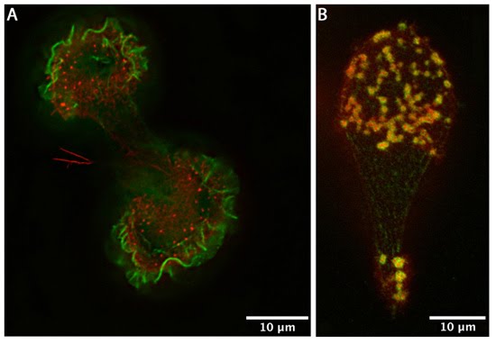
(A) Human acute monocytic leukemia cells (THP1) cultured on fibronectin, differentiated towards macrophages with TGF beta1, displaying podosomes (red puncta). Actin was visualized using phalloidin-TRITC. The image is a projection of several Z-planes where lamellipodial actin ruffles on upper planes are false-colored green and actin cores of podosomes on lower planes are inred. (B) Human acute monocytic leukemia cells (THP1) cultured on fibronectin, differentiated towards macrophages with TGF beta1, displaying podosomes (yellow puncta). Actin (red) was stained usingphalloidin-TRITC and vinculin (green) using Alexafluor488-coupled anti-vinculin antibody. The actin core and the surrounding ring complex (vinculin) are clearly visible. The cells were imaged using a DeltaVision microscope. [Images captured by Tee Yee Han, Mechanobiology Institute, Singapore]
Podosome structure
Structurally, the podosome is characterized by two main features – an actin core and a ring complex. The actin core contains several coordinators of actin nucleation, namely the Arp2/3 complex and WASP proximal to the plasma membrane and cortactin or HS-1 (hematopoietic lineage cell-specific protein 1) more distally [5]. Further to the dense actin network forming the core, actin filaments emanate radially from the actin core to the plasma membrane and between individual podosomes [6].
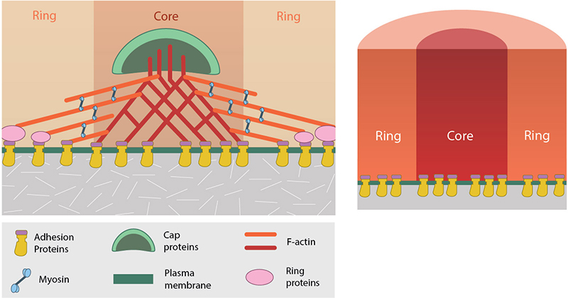
Podosomes have an actin-rich core region and a ring complex. Core actin binds to capping proteins whereas ring proteins bind to actin at the cell membrane.
The ring complex comprises integrins and integrin-associated proteins, such as paxillin [7] and serves to connect cell surface integrins with the cytoskeleton. Originally thought to be a round structure, recent advances in bioimaging have shown the ring complex to have a polygonal shape [8]. These findings were obtained by applying novel super-resolution analysis (Bayesian Blinking and Bleaching (3B) analysis) to data acquired by standard widefield microscopy of cells expressing fluorescently tagged proteins that localize to the podosome ring complex. The increased resolution of the images also suggested that different proteins within the ring complex have distinct localizations, with vinculin appearing more peripherally to talin [8].
In general, the podosome structure is no greater than approximately 0.5 μm in width and 1 μm in depth [7]. The lifetime of this structure is far shorter than that of focal adhesions, lasting only a few minutes [9].
References
- Rottiers P, Saltel F, Daubon T, Chaigne-Delalande B, Tridon V, Billottet C, Reuzeau E, and Génot E. TGFbeta-induced endothelial podosomes mediate basement membrane collagen degradation in arterial vessels. J. Cell. Sci. 2009; 122(Pt 23):4311-8. [PMID: 19887587]
- Calle Y, Burns S, Thrasher AJ, and Jones GE. The leukocyte podosome. Eur. J. Cell Biol. 2005; 85(3-4):151-7. [PMID: 16546557]
- Calle Y, Chou H, Thrasher AJ, and Jones GE. Wiskott-Aldrich syndrome protein and the cytoskeletal dynamics of dendritic cells. J. Pathol. 2004; 204(4):460-9. [PMID: 15495215]
- Murphy DA, and Courtneidge SA. The ‘ins’ and ‘outs’ of podosomes and invadopodia: characteristics, formation and function. Nat. Rev. Mol. Cell Biol. 2011; 12(7):413-26. [PMID: 21697900]
- Morton PE, and Parsons M. Dissecting cell adhesion architecture using advanced imaging techniques. Cell Adh Migr 2011; 5(4):351-9. [PMID: 21785274]
- Akisaka T, Yoshida H, Suzuki R, and Takama K. Adhesion structures and their cytoskeleton-membrane interactions at podosomes of osteoclasts in culture. Cell Tissue Res. 2007; 331(3):625-41. [PMID: 18087726]
- Linder S. The matrix corroded: podosomes and invadopodia in extracellular matrix degradation. Trends Cell Biol. 2007; 17(3):107-17. [PMID: 17275303]
- Cox S, Rosten E, Monypenny J, Jovanovic-Talisman T, Burnette DT, Lippincott-Schwartz J, Jones GE, and Heintzmann R. Bayesian localization microscopy reveals nanoscale podosome dynamics. Nat. Methods 2011; 9(2):195-200. [PMID: 22138825]
- Destaing O, Saltel F, Géminard J, Jurdic P, and Bard F. Podosomes display actin turnover and dynamic self-organization in osteoclasts expressing actin-green fluorescent protein. Mol. Biol. Cell 2003; 14(2):407-16. [PMID: 12589043]