
What are filopodia?
Filopodia (singular filopodium) are thin membrane protrusions that act as antennae for a cell to probe the surrounding environment [1][2][3]. Nonprotruding filopodia are mechanistically related to microspikes [4]. Filopodia are commonly found embedded within, or protruding from the lamelliopodium at the free front of migratory tissue sheets. Filopodia are also prominent in neurite growth cones and individual cells such as fibroblasts.
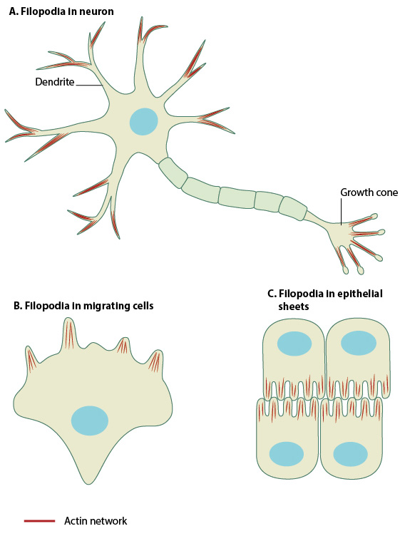
Filopodia are found in neurons (A), at the protruding edge in migrating cells (B), and in epithelial sheets (C).
Filopodia are 60-200 nm in diameter and contain parallel bundles of 10-30 actin filaments held together by actin-binding proteins (e.g. fascin). These filaments are oriented so that their barbed end is directed towards the protruding membrane.
Filopodia sense the extracellular environment at their tips using cell surface receptors [5][6][7]. Contact with an external target promotes the coupling of membrane-bound proteins to the backward (retrograde) flow of actin; this coupling produces the pulling forces needed for cell migration processes such as wound healing and neurite growth [8]. Contact differences between substrates or cell types influences the number of protruding filopodia [9].
A key set of proteins is involved in filopodia formation; however, the relative importance of each protein seems to vary between different organisms and their cell types. Three basic steps are involved in filopodial assembly: filament nucleation, sustained barbed end elongation and filament bundling.
Steps in Filopodia Formation and Function
Filopodia are dynamic structures that are primarily composed of F-actin bundles and whose initiation and elongation are precisely regulated by the rate of actin filament assembly, convergence and cross-linking.
The actin treadmilling mechanism of elongation is essential in protrusion [10] and any change in the frequency of initiation, or the balance of extension versus retraction of actin filaments, can result in the gain or loss of filopodia. Filopodial actin filaments are unbranched [11], and observations of filopodial formation have revealed that actin assembles at the filopodial tips, moves backwards, and dissipates at the rear [12].
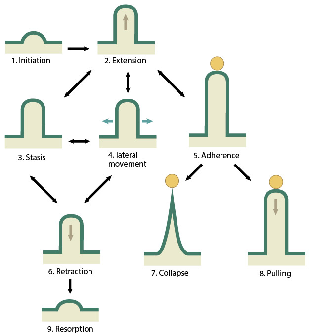
Filopodia undergo 9 distinct steps in their formation. Double-sided arrows symbolize the ability of a filopodium to oscillate between different states.
References
- Davenport RW, Dou P, Rehder V, and Kater SB. A sensory role for neuronal growth cone filopodia. Nature 1993; 361(6414):721-4. [PMID: 8441465]
- Mattila PK, and Lappalainen P. Filopodia: molecular architecture and cellular functions. Nat. Rev. Mol. Cell Biol. 2008; 9(6):446-54. [PMID: 18464790]
- Yuan X, Jin M, Xu X, Song Y, Wu C, Poo M, and Duan S. Signalling and crosstalk of Rho GTPases in mediating axon guidance. Nat. Cell Biol. 2003; 5(1):38-45. [PMID: 12510192]
- Svitkina TM, Bulanova EA, Chaga OY, Vignjevic DM, Kojima S, Vasiliev JM, and Borisy GG. Mechanism of filopodia initiation by reorganization of a dendritic network. J. Cell Biol. 2003; 160(3):409-21. [PMID: 12566431]
- Sheetz MP, Baumrind NL, Wayne DB, and Pearlman AL. Concentration of membrane antigens by forward transport and trapping in neuronal growth cones. Cell 1990; 61(2):231-41. [PMID: 2331749]
- Letourneau PC, and Shattuck TA. Distribution and possible interactions of actin-associated proteins and cell adhesion molecules of nerve growth cones. Development 1989; 105(3):505-19. [PMID: 2612362]
- Steketee MB, and Tosney KW. Three functionally distinct adhesions in filopodia: shaft adhesions control lamellar extension. J. Neurosci. 2002; 22(18):8071-83. [PMID: 12223561]
- Lamoureux P, Buxbaum RE, and Heidemann SR. Direct evidence that growth cones pull. Nature 1989; 340(6229):159-62. [PMID: 2739738]
- Bastmeyer M, and Stuermer CA. Behavior of fish retinal growth cones encountering chick caudal tectal membranes: a time-lapse study on growth cone collapse. J. Neurobiol. 1993; 24(1):37-50. [PMID: 8419523]
- Small JV. Lamellipodia architecture: actin filament turnover and the lateral flow of actin filaments during motility. Semin. Cell Biol. 1994; 5(3):157-63. [PMID: 7919229]
- Ahmed S, Goh WI, and Bu W. I-BAR domains, IRSp53 and filopodium formation. Semin. Cell Dev. Biol. 2009; 21(4):350-6. [PMID: 19913105]
- Mallavarapu A, and Mitchison T. Regulated actin cytoskeleton assembly at filopodium tips controls their extension and retraction. J. Cell Biol. 1999; 146(5):1097-106. [PMID: 10477762]