
How are intermediate filaments assembled?
The soluble subunit for creating intermediate filaments is a tetramer. The tetramer is created from monomers in a stepwise fashion (as reviewed in [1]). First, two monomers associate via their central domains to form parallel helical coils around each other. This parallel dimer then associates with another parallel dimer in an antiparallel arrangement to form a staggered tetramer. The lateral association of eight tetramers results in the formation of a unit-length filaments (ULF). Two ULFs are able to anneal in an end-to-end fashion (i.e. longitudinally) to form a thick filament, approximately 16 nm in diameter. Further end-to-end annealing of ULFs results in filament elongation, which is followed by radial compaction to achieve the final intermediate filament diameter [2][3].
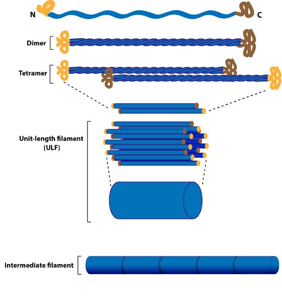
Intermediate filaments are built from monomers that associate with each other form dimers. Pairs of dimers then associate in an anti-parallel fashion to form staggered tetramers. Lateral associations between eight tetramers form unit-length filaments, which are able to anneal to each other, end-to-end, to form intermediate filaments.
What are some unique features of intermediate filaments?
IFs are heterogenous
When compared to actin and tubulin, there is greater sequence variation within intermediate filament genes and the proteins they produce. This yields a greater diversity in the types of structures they can form (e.g. hair, nails, feathers, horns). Although different intermediate protein classes share a common ancestral origin, intermediate filament genes are only conserved in metazoans. Fungi and plants lack these filaments and insects have only one class [4]. It seems likely that as the metazoan lineage evolved more intricate and complex tissues and organs, intermediate filaments were adapted and modified for tissue or cell specific purposes.
IFs lack polarity
Intermediate filament subunits do not bind nucleotides and are oriented in opposite directions during filament assembly. This leads to a similar structural appearance at both the filament ends, as suggested by FRAP (fluorescence recovery after photobleaching) experiments [5]. Furthermore subunit exchange is not confined to filament ends, as with microtubules and actin filaments, but occurs along the length of the filament [6].
IFs have a high tensile strength and are resistant to compression, twisting and bending forces
The elastic nature of intermediate filaments is due the staggered assembly of their subunits into protofilaments and the high degree of lateral versus longitudinal interactions within the filaments. This structure promotes high tensile strength, toughness and long-range elasticity [7](reviewed in [8]).
IFs lack motor proteins
No motor proteins have been identified to move along intermediate filaments as they lack polarity [9].
IFs have slower filament dynamics
IFs exhibit a dynamic exchange of their constituent subunits, however this exchange rate is much slower than those observed with actin filaments or microtubules [6].
References
- Herrmann H, Strelkov SV, Burkhard P, and Aebi U. Intermediate filaments: primary determinants of cell architecture and plasticity. J. Clin. Invest. 2009; 119(7):1772-83. [PMID: 19587452]
- Colakoğlu G, and Brown A. Intermediate filaments exchange subunits along their length and elongate by end-to-end annealing. J. Cell Biol. 2009; 185(5):769-77. [PMID: 19468066]
- Kirmse R, Portet S, Mücke N, Aebi U, Herrmann H, and Langowski J. A quantitative kinetic model for the in vitro assembly of intermediate filaments from tetrameric vimentin. J. Biol. Chem. 2007; 282(25):18563-72. [PMID: 17403663]
- Mencarelli C, Ciolfi S, Caroti D, Lupetti P, and Dallai R. Isomin: a novel cytoplasmic intermediate filament protein from an arthropod species. BMC Biol. 2011; 9:17. [PMID: 21356109]
- Vikstrom KL, Lim SS, Goldman RD, and Borisy GG. Steady state dynamics of intermediate filament networks. J. Cell Biol. 1992; 118(1):121-9. [PMID: 1618899]
- Colakoğlu G, and Brown A. Intermediate filaments exchange subunits along their length and elongate by end-to-end annealing. J. Cell Biol. 2009; 185(5):769-77. [PMID: 19468066]
- Fudge DS, Gardner KH, Forsyth VT, Riekel C, and Gosline JM. The mechanical properties of hydrated intermediate filaments: insights from hagfish slime threads. Biophys. J. 2003; 85(3):2015-27. [PMID: 12944314]
- Kreplak L, and Fudge D. Biomechanical properties of intermediate filaments: from tissues to single filaments and back. Bioessays 2007; 29(1):26-35. [PMID: 17187357]
- Herrmann H, Strelkov SV, Burkhard P, and Aebi U. Intermediate filaments: primary determinants of cell architecture and plasticity. J. Clin. Invest. 2009; 119(7):1772-83. [PMID: 19587452]